


:strip_icc()/s03.video.glbimg.com/x720/12610546.jpg)
The scan in mice has a resolution up to 64 million times better than brain MRI techniques currently used.
1 hour ago
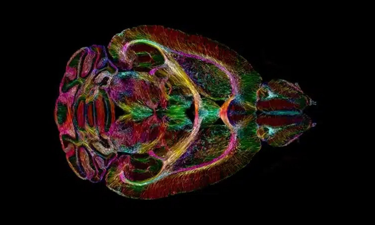
In 1973, scientists performed the first magnetic resonance imaging in history. Fifty years have passed, and now a team of researchers from the Center for In Vivo Microscopy at Duke University, in the United States, has achieved the clearest picture of the brain.
Test performed with an accuracy of up to 64 million times better Brain MRI techniques used today. The full study is available in the scientific journal PNAS.
The brain shown in the image belongs to a mouse. Analyzing an animal’s organ will help scientists understand issues that also affect humans, such as changes in the brain with age, the influence of diet and even the emergence of neurodegenerative diseases such as Alzheimer’s.
The picture did not appear overnight. In fact, it is the fruit of four decades of study.
The team used, for example, a 9.4 Tesla magnet (a unit used for magnetic flux density). In clinical MRI, magnets from 1.5 to 3 Tesla are usually used. A special set of graded coils 100 times more powerful than conventional coils and a computer with the performance of 800 laptops were used.
The scientists also applied a technique known as optical sheet microscopy. Basically, it allows you to classify specific groups of cells in the brain, such as dopamine-emitting cells, to look at the development of diseases like Parkinson’s.
The study showed how brain connectivity changes as the mice age, as well as significant changes in specific regions. A set of images indicates the marked deterioration of neural networks in a mouse model of Alzheimer’s disease.

“Incurable thinker. Food aficionado. Subtly charming alcohol scholar. Pop culture advocate.”
More Stories
Games from the Kingdom Hearts series are finally coming to Steam
Your cell phone battery doesn’t last because of these three useless functions
Black Myth Wukong can have 80 bosses and 160 enemy types